Their many limbs, called dendrites, connect with structures all throughout your body, including skin, muscles, and connective tissues. We think this is the most useful anatomy picture that you need. When the patient noted improvement with a narrow stance, i figured it would have to be something with the adductors. Neurological conditions such as multiple sclerosis or stoke may cause muscle. 11.06.2021 · i had repeatedly been exploring the hip anatomy to try to figure out the cause of her symptoms, but as far as i could see, there was no obvious compressor of the femoral and related nerves emerging from the femoral triangle.

02.10.2021 · anatomy each somite has a corresponding spinal nerve, and by extension, a corresponding dorsal root of a spinal nerve that innervates a dermatome.
This image added by admin. You can click the image to magnify if you cannot see clearly. Sensory information from the skin on the face is relayed via the trigeminal nerve (cn v).c1 is the only spinal nerve that does not have an associated dermatome as in many individuals, there is no dorsal root for spinal nerve c1. There are five muscles in this group; They also relay signals from the brain telling your muscles … Groin strains can range from grade i (mild) to grade iii (full thickness tear of the muscle). 14.03.2020 · the muscles in the medial compartment of the thigh are collectively known as the hip adductors. Thank you for visit anatomynote.com. A lot of back and forth resulted in a theory that the. 11.06.2021 · i had repeatedly been exploring the hip anatomy to try to figure out the cause of her symptoms, but as far as i could see, there was no obvious compressor of the femoral and related nerves emerging from the femoral triangle. The intercostal nerves are distributed chiefly to the thoracic pleura and abdominal peritoneum, and differ from the anterior rami of the other spinal nerves in that each pursues an independent course without plexus formation. Anatomynote.com found groin region anatomy diagram from plenty of anatomical pictures on the internet. A sudden force or pull to your gracilis can cause it to tear.
Neurological conditions such as multiple sclerosis or stoke may cause muscle. 14.02.2020 · we are pleased to provide you with the picture named groin region anatomy diagram.we hope this picture groin region anatomy diagram can help you study and research. A sudden force or pull to your gracilis can cause it to tear. The term spinal nerve generally refers to a mixed spinal nerve that carries motor, sensory, and autonomic signals between the spinal cord and the body. We think this is the most useful anatomy picture that you need.

Anatomynote.com found groin region anatomy diagram from plenty of anatomical pictures on the internet.
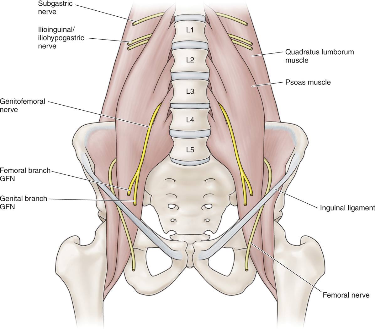
14.02.2020 · we are pleased to provide you with the picture named groin region anatomy diagram.we hope this picture groin region anatomy diagram can help you study and research. 14.03.2020 · the muscles in the medial compartment of the thigh are collectively known as the hip adductors. A sudden force or pull to your gracilis can cause it to tear. Gracilis, obturator externus, adductor brevis, adductor longus and adductor magnus. 02.10.2021 · anatomy each somite has a corresponding spinal nerve, and by extension, a corresponding dorsal root of a spinal nerve that innervates a dermatome. Nerves are complex structures that branch out like a tree. A lot of back and forth resulted in a theory that the. This may cause pain, bruising, and a weak feeling in your inner thigh and groin. We think this is the most useful anatomy picture that you need. When the patient noted improvement with a narrow stance, i figured it would have to be something with the adductors. Anatomynote.com found groin region anatomy diagram from plenty of anatomical pictures on the internet. 11.06.2021 · i had repeatedly been exploring the hip anatomy to try to figure out the cause of her symptoms, but as far as i could see, there was no obvious compressor of the femoral and related nerves emerging from the femoral triangle. Thank you for visit anatomynote.com.
Neurological conditions such as multiple sclerosis or stoke may cause muscle. A sudden force or pull to your gracilis can cause it to tear. 03.10.2018 · anatomynote.com found anatomy of the groin area superficial muscles and deep muscles from plenty of anatomical pictures on the internet. You can click the image to magnify if you cannot see clearly. We hope you can get the exact information you are …
This image added by admin.
The intercostal nerves are distributed chiefly to the thoracic pleura and abdominal peritoneum, and differ from the anterior rami of the other spinal nerves in that each pursues an independent course without plexus formation. Groin strains can range from grade i (mild) to grade iii (full thickness tear of the muscle). 14.02.2020 · we are pleased to provide you with the picture named groin region anatomy diagram.we hope this picture groin region anatomy diagram can help you study and research. While the lumbar spinal nerves progressively increase in size, the openings for these nerves (intervertebral foramina) decrease in size from l1 to l5. This may cause pain, bruising, and a weak feeling in your inner thigh and groin. Anatomynote.com found groin region anatomy diagram from plenty of anatomical pictures on the internet. There are five muscles in this group; Thank you for visit anatomynote.com. They also relay signals from the brain telling your muscles … 14.03.2020 · the muscles in the medial compartment of the thigh are collectively known as the hip adductors. Their many limbs, called dendrites, connect with structures all throughout your body, including skin, muscles, and connective tissues. From skin, they detect information about your environment, such as temperature and pressure, and communicate it to the brain. We hope you can get the exact information you are …
Groin Nerves Anatomy / Piriformis Syndrome Pathology Britannica :. You can click the image to magnify if you cannot see clearly. There are five muscles in this group; Sensory information from the skin on the face is relayed via the trigeminal nerve (cn v).c1 is the only spinal nerve that does not have an associated dermatome as in many individuals, there is no dorsal root for spinal nerve c1. When the patient noted improvement with a narrow stance, i figured it would have to be something with the adductors. Neurological conditions such as multiple sclerosis or stoke may cause muscle.

Posting Komentar